Figure 2.
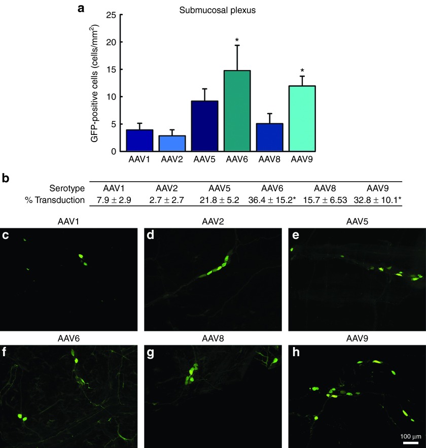
Transduction efficiency of recombinant adeno-associated virus (AAV) in the submucosal plexus (SMP) of the enteric nervous system: a comparison across serotypes 1, 2, 5, 6, 8, and 9. Adult male rats received direct injections of AAV (6 × 5 µl injections at 1.3 × 10¹² vg/ml) into the wall of the descending colon and were sacrificed one month later, at which time the SMP was dissected and viewed under a fluorescent microscope. (a) Transduction efficiency of AAV1, 2, 5, 6, 8, and 9 was measured by the quantification of green fluorescent protein (GFP) expressing cells in the SMP. Columns represent mean number of GFP-positive cells, expressed as cells/mm2, + 1 SEM (n = 4–6/group). AAV6 and 9 exhibited significantly greater numbers of GFP-positive cells than AAV1 and AAV2. (b) The percent of HUc-positive cells transduced for each respective serotype. Table represents the percent of HUc cells colocalized with GFP ± SEM (n = 4–6/group). (c–h) Representative images of GFP-positive cells in the SMP following transduction by AAV1, 2, 5, 8, and 9. Scale bar in (h) represents 100 µm and applies to panels (c–g). * Indicates significantly different than AAV1 and AAV2 (P < 0.05).

