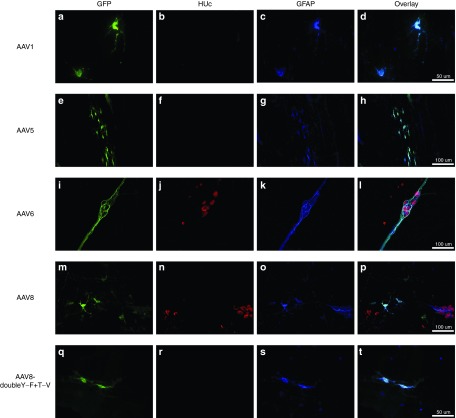
Figure 5.
Glial tropism of recombinant adeno-associated virus (AAV) transduction. Adult male rats received direct injections of AAV (6 × 5 µl injections at 1.3 × 10¹² vg/ml) into the wall of the descending colon and were sacrificed one month later, at which time the submucosal plexus (SMP) and myenteric plexus (MP) were dissected. MP and SMP tissue was stained for the enteric glia marker, glial fibrillary acidic protein (GFAP; blue). Following, tissue was viewed under a fluorescent microscope and the transduction of glia was determined by examining the colocalization of green fluorescent protein (GFP; green) expression in GFAP-positive enteric glia (colocalization appears turquoise). Micrographs also show the neuronal marker HUc (red). Glia transduction was rare and was only observed in AAV serotypes 1, 5, 6, 8 and AAV8-doubleY-F+T-V. AAV6 showed far more glial transduction than any other serotype. Morphologically, most glial transduction appeared to occur within reactive, protoplasmic or mucosal, enteric glia (d,h,p, and t). In contrast, AAV6 was able to transduce intraganglionic enteric glia (l). Representative images of glial transduction are shown for AAV1 in the MP (a–d), AAV5 in the MP (e–h), AAV6 in the SMP (i–l), AAV8 in the SMP (m–p), and AAV8-doubleY-F+T-V in the SMP (q–t). Scale bar in (d,t) represents 50 µm and applies to (a–c) and (q–s). Scale bars in (h,l,p) represent 100 µm and apply to (e–g,i–k,m–o).

