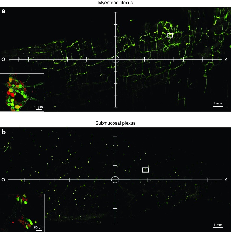
Figure 7.
The spread of a single injection of recombinant adeno-associated virus (AAV) serotype 9 in the myenteric plexus (MP) and submucosal plexus (SMP) of the enteric nervous system. Adult male rats received direct injections of AAV9 (1.2 × 10¹³ vg/ml) into the wall of the descending colon and were sacrificed one month later, at which time the MP and SMP were dissected and viewed under a fluorescent microscope. The spread of transduction was determined by quantifying the area of the MP and SMP containing green fluorescent protein (GFP)-positive cells. Panel (a) and (b) show representative images of vector spread and transduction following a single injection of AAV9 within the MP and SMP, respectively. Representative images are shown with axis to indicate longitudinal (x-axis) and perpendicular (y-axis) spread. Tick marks on the axis represent 1 mm. The circle in the center of the axis represents the approximate location of the injection site. Insets represent high magnification images of the area within the white box in a and b. Scale bars in (a) and (b) represent 1 mm. Scale bars in the insets in (a) and (b) represent 50 μm.

