. 2015 Feb 28;39(1):108–115. doi: 10.5535/arm.2015.39.1.108

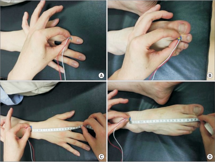
Fig. 2. Stimulation site of pain-related evoked potential. (A) In upper limb stimulation, the stimulating electrode was placed on the middle phalanx of the second digit of the hand (C7 dermatome). (B) In lower limb stimulation, the stimulating electrode was placed on the middle phalanx of the second toe of the foot (L5 dermatome). (C) For calculating upper extremity conduction velocity, distal N latency was measured by stimulating the middle phalanx of the second digit. Proximal N latency was measured by stimulating the point where it measured from 20 cm proximal from the middle phalanx of the second digit. The 20-cm distance between distal and proximal stimulation sites was divided by the difference between the distal N latency and the proximal N latency. (D) The same method was used for calculating lower extremity conduction velocity.

Fig. 2