Figure 2.
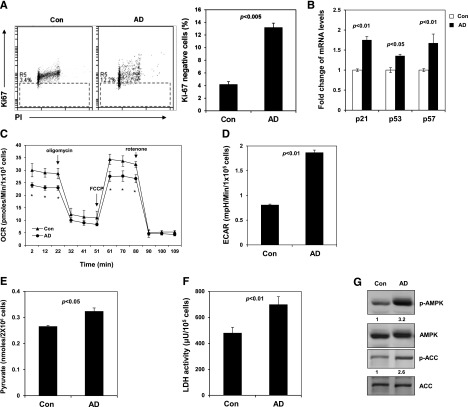
Inhibition of Ptpmt1 maintains stem cell quiescence by reprogramming cellular metabolism and activation of AMPK. (A) Sorted LSK cells were cultured as described in Figure 1C. After 7 days, cells were collected, fixed, and stained with fluorescein isothiocyanate –labeled anti-Ki67 antibody and propidium iodide (PI). Percentage of Ki67-negative cells (quiescent cells) was quantified by FACS. (B) Messenger (m)RNA levels of p21, p57, and p53 in cultured LSK cells were determined by quantitative reverse-transcription polymerase chain reaction. (C-D) Sorted LSK cells were treated with AD or vehicle as in panel A. Oxygen consumption rate (OCR) (C) and extracellular acidification rates (ECAR) (D) were measured. Experiments were repeated 3 times, and similar results were obtained in each. Representative results from 1 experiment are shown; *P < .05. (E) Lin− cells were cultured in the presence of AD (250 nM) or vehicle for 24 hours. Intracellular pyruvate concentration was determined using the pyruvate assay kit (Cayman Chemical Company, Ann Arbor, MI). (F) Intracellular lactate dehydrogenase (LDH) activity was determined using the LDH assay kit (Cayman Chemical Company). (G) Sorted LSK cells were treated with AD or vehicle as in panel A. Whole-cell lysates were prepared and examined by western blot analysis with anti-phospho-AMPK (Thr172) and anti-phospho-ACC (Ser79) antibodies. Blots were stripped and reprobed with anti-AMPK and anti-ACC antibodies. Relative phospho- (p-)AMPK and p-ACC levels normalized against pan protein levels are shown. Representative results from 3 independent experiments are shown. ACC, acetyl-CoA carboxylase.

