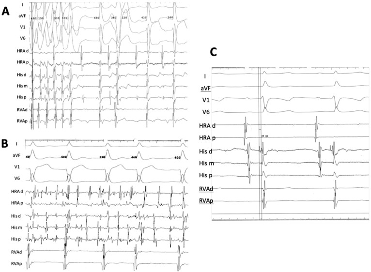
Figure 4. Electrophysiological Study Tracings of Two Patients with NKX2-5 Truncating Mutations.
Multipolar electrode catheters were introduced percutaneously from the right femoral vein and positioned into the high right atrium (HRA) right ventricular apex (RVA) and His bundle region of two patients who carry NKx2.5 mutations. Bipolar electrograms (30 to 500 Hz) were displayed and stored using a digital recording system (EP MedSystems, West Berlin, NJ). One of the patients (B II:6) had no baseline ventriculo-atrial (VA) conduction, an atrial tachycardia that degenerated into atrial fibrillation requiring DC cardioversion (B), and spontaneous premature ventricular complexes isolated and in couplets with left bundle inferior axis morphology with early precordial transition (A). The other patient (B III:1) was in normal sinus rhythm, had a normal atrial-His (AH) interval, a short His-ventricular (HV) interval at 26 ms (C) [Normal HV interval: 35–55 ms] and no VA conduction was observed with or without isoproterenol infusion.

