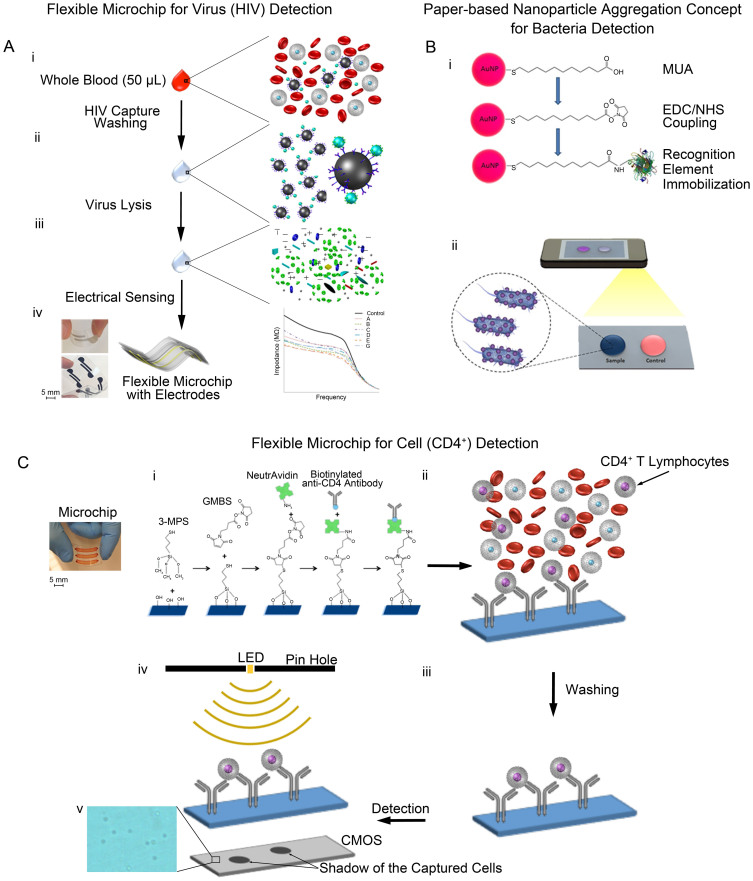
Figure 1. Schematic of seamlessly integrated paper and flexible substrate-based platforms with broadly applicable electrical and optical sensing modalities.
(A) Flexible electrodes on polyester film-based electrical sensing platform for HIV detection. (i) Viruses are captured using streptavidin-coated magnetic beads conjugated with biotinylated polyclonal anti-gp120 antibodies in clinically relevant samples such as whole blood (ii) Sample is washed to remove electrically conductive solution. (iii) Captured viruses are lysed using 1% Triton X-100, releasing to release ions and biomolecules of the captured viruses. These biomolecules change the electrical conductivity of the solution. (iv) The impedance magnitude of the viral lysate samples decreases compared with that of control samples. Figure 1A is drawn by H.S. (B) Nanoparticle aggregation concept for bacteria detection on a cellulose paper and its incorporation with mobile phone camera. (i) Gold nanoparticles (AuNPs) are modified with 11-Mercaptoundeconoic acid (MUA), N-Ethyl-N'-(3-dimethylaminopropyl) carbodiimide hydrochloride (EDC), and N-hydroxysuccinimide (NHS) to form coupling groups on nanoparticle surface. AuNPs are further functionalized with specific recognition elements for bacteria pathogens. (ii) Bacteria-spiked samples are added to the modified nanoparticle solution. Bacteria samples cause nanoparticle aggregation and change the color of solution as detected with a mobile phone. Figure 1B is drawn by F.I. (C) Lensless imaging detection and counting of CD4+ T lymphocytes on polyester film-based platform with microchannels. (i) Surface chemistry on the platform are designed to immobilize biotinylated anti-CD4 antibodies using NeutrAvidin on chemically activated surface including N-g-Maleimidobutyryloxy succinimide ester (GMBS) and 3-Mercaptopropyl trimethoxysilane (3-MPS) modifications. (ii) CD4+ T lymphocytes are selectively captured from a whole blood sample on the surface of platform. (iii) Non-captured cells are washed away from the microchannels of platform. (iv) Captured CD4+ T cells are then detected and quantified using a lensless shadow imaging technology. (v) Shadows of captured CD4+ T cells on the substrate. Figure 1C is drawn and designed by H.S., W.A. and M.H.Z.

