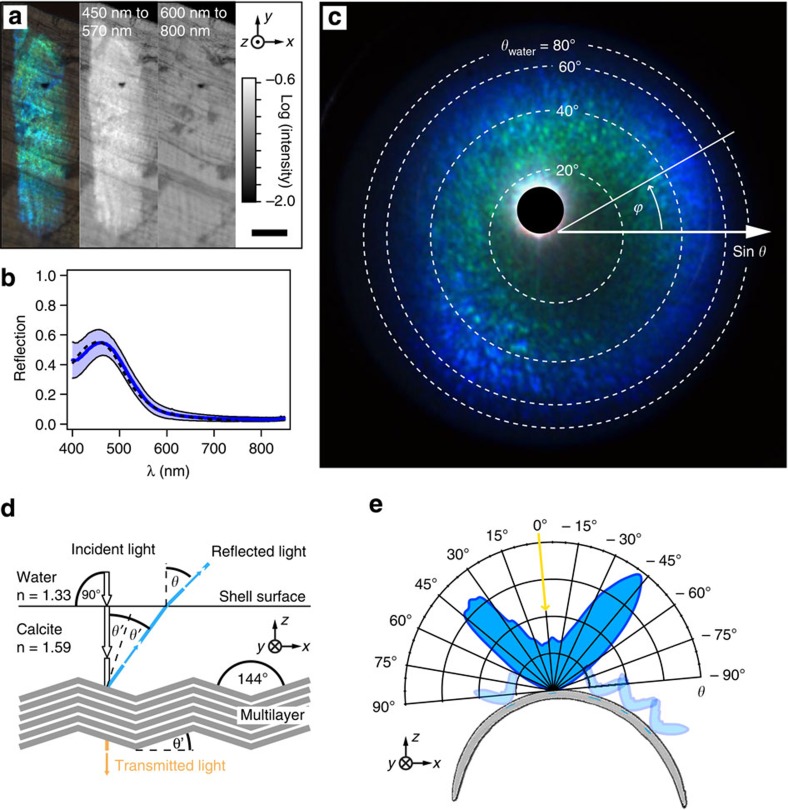
Figure 4. Optical properties of the photonic multilayer.
(a) Reflection optical micrograph of a single stripe (left) in comparison with the two intensity maps of light reflected in the range of 450–570 nm (middle) and 600–800 nm (right) wavelengths. Scale bar, 50 μm. (b) Reflection spectrum of a stripe in water, referenced against a 97% reflective silver mirror. The blue line represents the average reflectivity with the s.d. being visualized by the blue shaded area (seven limpets, 10–20 measurements per shell). The black dashed curve represents theoretical calculations of the blue stripes’ reflectivity that results from 500 calculation runs assuming a multilayer stack with 40 calcite lamellae (n=1.59) and water filling the interstitial gaps (n=1.33) with a lamellae thickness of 113±11 nm and a spacing of 53±7 nm determined from TEM images. (c) Diffraction microscopy image visualizing the scattering upon light reflection from a single stripe in water, where each pixel represents the intensity of light as a function of its propagation direction after reflecting off the stripe as characterized by the polar angle θ (measured from the shell surface normal) and the azimuthal angle ϕ. The blanked bright spot is an artifact caused by non-avoidable internal reflections in the microscope setup. (d) Schematic representation of light reflection from the limpet’s photonic zig-zag multilayer structure for light normally incident on the shell surface. According to Snell’s law, the angle for which the highest reflectivity of a stripe in water is observed, θ≈40°, corresponds to an angle of ≈32° (with respect to the incident light) in the calcite shell suggesting an inclination of the reflecting multilayer of θ’≈16° with respect to the shell surface, which matches well with the observed characteristic angle of around 144° between the multilayer lamellae. (e) Polar plot of reflection intensity showing that each stripe reflects light in a broad angular range of more than a 60° cone angle. This data was obtained by averaging cross-sections of the data shown in c at different azimuthal angles ϕ. Due to the different orientations of the stripes along the curved shell, portions of the stripe pattern can be clearly seen from almost any direction.

