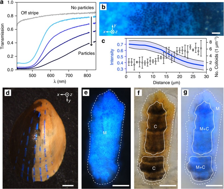
Figure 6. Optical function of the particles beneath the multilayer.
(a) Light transmission along a path from particle-free zones to particle-covered regions beneath a blue stripe. (b) High-resolution optical micrograph acquired in reflection of the transition area from the edge of a blue stripe (no particles, left) towards the centre (dense particle coverage, right). Scale bar, 2 μm. (c) Correlation of transmitted light intensity averaged over the wavelength range of 400–750 nm and particle density deduced from SEM cross-sections along a path from left to right of b. (d) Photograph of a limpet shell with its right interior side painted white. Scale bar, 1 mm. (e,f) Reflection and transmission optical image of a single stripe before paint application. The circumference of the stripe, that is, the area where the multilayer (M) is located, is marked with a white dashed line. The locations of colloidal particles (C) beneath the multilayer are marked with white solid lines. (g) Reflection optical image of the same stripe after painting. The areas with multilayer and colloidal particles (M+C) maintain a strong blue coloration while areas where particles are missing below the multilayer (M) display only a faint blue colour. Scale bars in (e–g), 50 μm.

