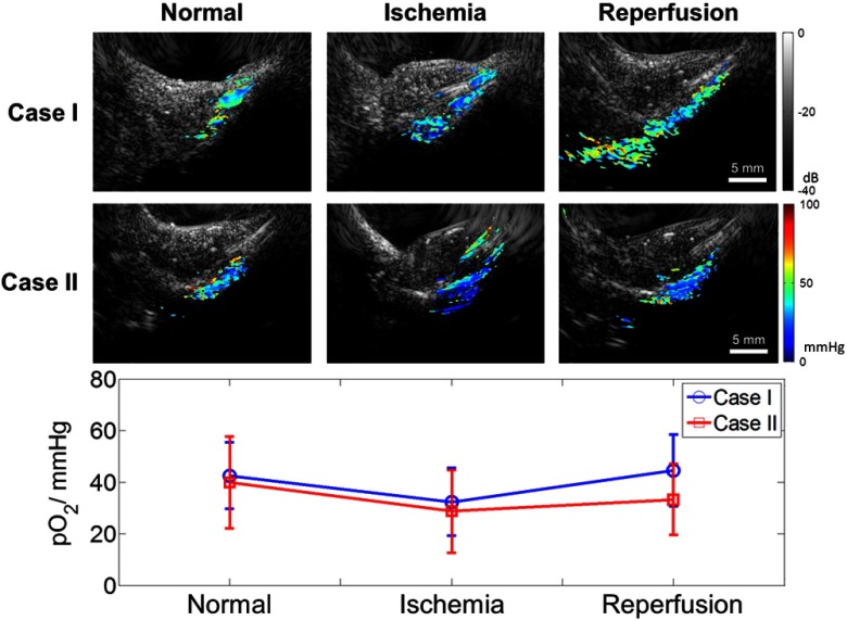
Fig. 4.
PALI tracking of during a blood-flow-restricting experiment in the upper part of hindlimb of normal mice. Combined US/PALI images are shown before (column under normal), during flow restriction (ischemia), and immediately after releasing the restriction (reperfusion). Cases I and II were experiments done on two mice. Statistics of the pixel values imaged are given by the average and error bars as standard deviation.

