
Figure 5.
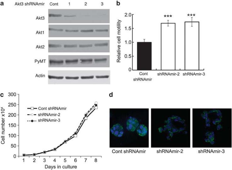
Knockdown of Akt3 increases cell migration without affecting cell growth. It also disrupts mammary acinar structure. (a) Three different Akt3 shRNAmirs targeting different regions of the Akt3 mRNA and non-silencing vector control were transduced into a PyMT mammary tumor cell line. The cells were lysed and western blotted with antibodies against Akt3, Akt2, Akt1 and Actin. (b) The motility of PyMT cell lines transduced with either control shRNAmir, Akt3 shRNAmir-2 or Akt3 shRNAmir-3 was examined by transwell migration assays. The number of migrated cells in each well was counted from seven × 20 microscope fields. The bar graphs show the average number of migrated cells per field±s.e.m. from three independent experiments performed in triplicates; ***P<0.001, by two-tailed t-test. (c) Cell growth curves of PyMT cell lines transduced with Akt3 shRNAmir-2 or -3 were compared with that of cells expressing control shRNAmir. Each time point represents mean±s.e.m. of cell numbers from triplicate experiments. (d) PyMT cell lines transduced with either control shRNAmir, Akt3 shRNAmir-2 or -3 were grown in Matrigel for 10 days. Cultures were fixed and stained with phalloidin and counterstained with 4,6-diamidino-2-phenylindole (DAPI). For each condition, acini formation was imaged by confocal microscopy and representative images are shown.