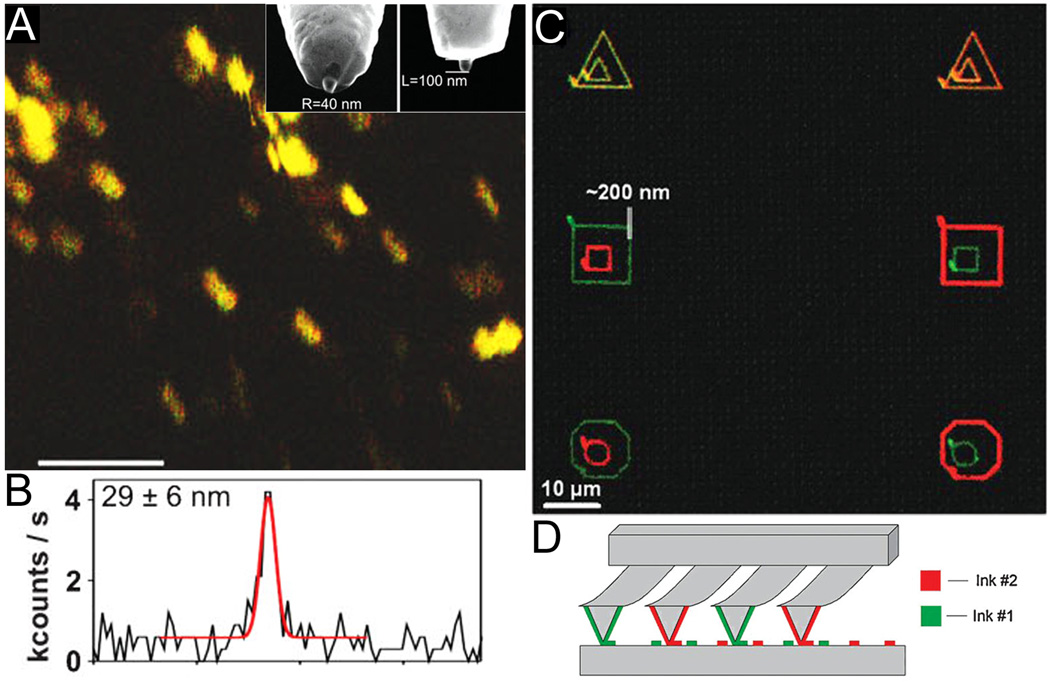
Figure 2.
Scanning probes have been used both for nanoscale imaging and patterning of membranes. (A) Fluorescence enhancement at the tip of nano-antenna on a scanning probe provides 30 nm resolution of membrane-bound integrin LFA-1; scale bar = 1 µm. A SEM image of the nano-antenna is shown in the inset of (A). (B) A line scan of fluorophore emission intensity vs. distance demonstrates 29 nm resolution. (C) SLBs of varying composition have been simultaneously patterned with dip-pen lithography at 200 nm resolution. (D) Multiplexed “pens” with varying composition of lipid-containing inks allow for simultaneous writing of many patterns and combinatorial mixing of the membrane components. (Reprinted with permission from (A,B) Ref. 101 and (C,D) Ref. 78.)

