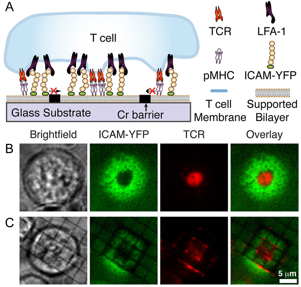
Figure 3.
Membrane reorganization is induced upon the binding of T cells to antigen presenting cells, forming an immunological synapse. (A) Key elements of antigen presenting cells (i.e. the peptide-major histocompatiability complex (pMHC) and the inter-cellular adhesion molecule (ICAM)) have been incorporated into a SLB and reorganized upon binding to T cell receptors (TCR) and to lymphocyte function-associated antigen 1 (LFA-1), respectively. The (B) presence or (C) absence of the metallic barriers to diffusion results in distinctly different morphology of the resulting immunological synapse as the T cell attempts to concentrate the TCRs within a ring of ICAMs. (Reprinted with permission from Ref. 32.)

