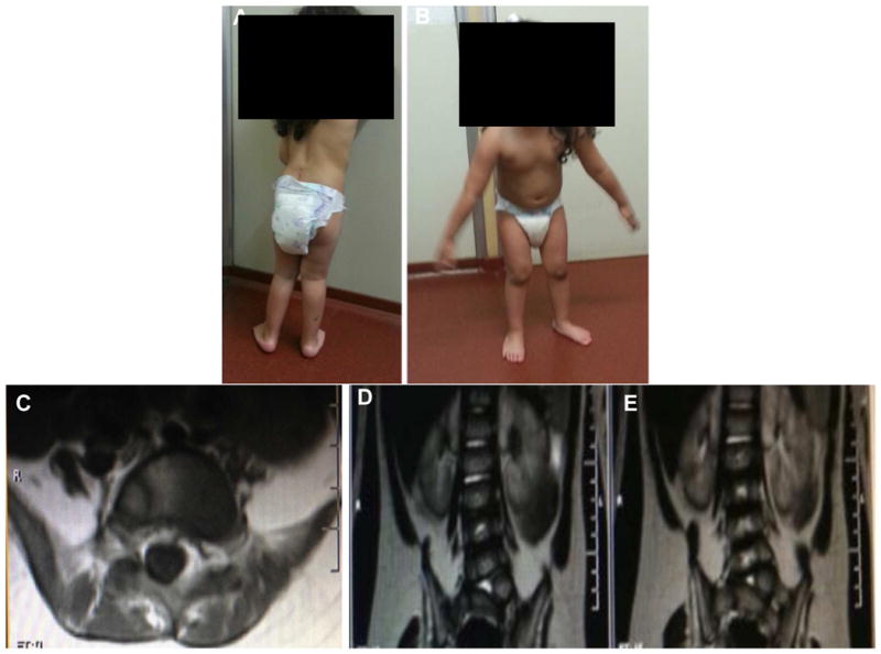
Figure 1.

Patient at 3.6 years of age. Panel A: Patient in standing position showed left scoliosis and calf atrophy. Panel B: Patient in standing position showed selective atrophy of lower extremities. Panel C: Coronal and Panel D: Axial lumbarsacral spine MRI of patient (long and short arrow respectively) showed left vertebral anomaly (hemivertebra) corresponding to level L5/S1.
