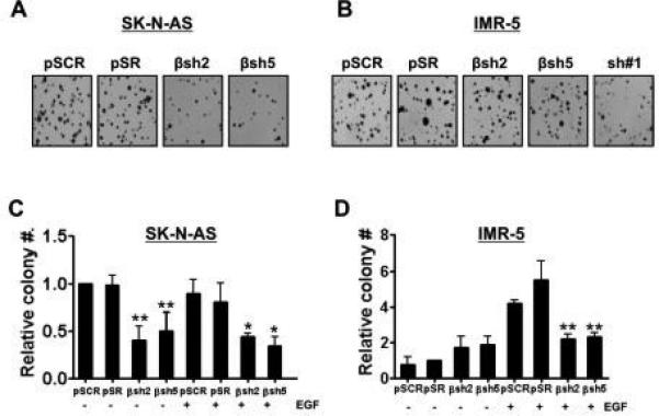
Figure 2. PI3KC2β-silencing decreases anchorage-independent growth of IMR-5 NB cells.

SK-N-AS (A) and IMR-5 (B) NB cells expressing the indicated shRNAs were plated in triplicate in soft agar. Colonies were stained with MTT after 3 weeks of growth and then photographed using a digital scanner. SK-N-AS (C) and IMR-5 (D) cells infected with indicated shRNAs or controls were plated in triplicate in soft agar with or without 100 ng/ml of EGF. Colonies were stained with MTT after 3 weeks of growth and then photographed using a digital scanner. Colonies were counted using the Image Quant LAS4010. Data represent the mean +/− standard error of three independent experiments. For SK-N-AS, βsh2 p=0.0026** and βsh5 p=0.0072** compared to control pSCR without EGF; for EGF-treatment, βsh2 p=0.021* and βsh5 p=0.029* compared to control pSCR. For IMR-5 in absence of EGF stimulation, differences observed with βsh2 and βsh5 were not significant compared to control pSCR. For EGF-treated samples, βsh2 p=0.00029** and βsh5 p=0.00022** compared to control pSCR.
