Figure 6.

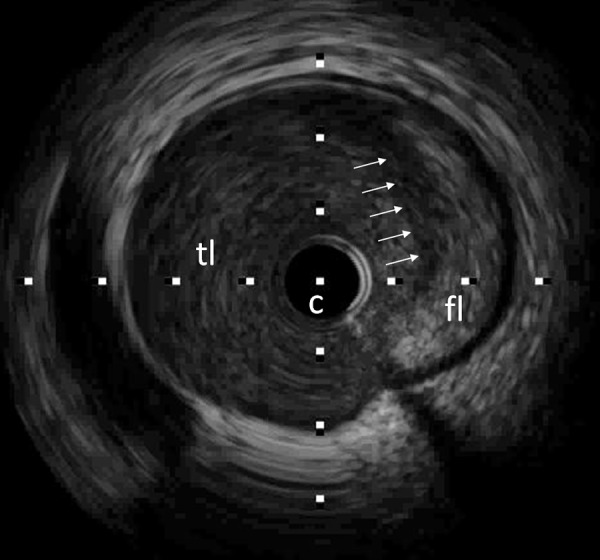
Intracoronary ultrasound (IVUS) of the corresponding LAD segment demonstrates a false lumen (fl) occupied by an echogenic mass (intramural hematoma). The true lumen (tl) is compressed and narrowed (white arrows: flap, c: catheter).

Intracoronary ultrasound (IVUS) of the corresponding LAD segment demonstrates a false lumen (fl) occupied by an echogenic mass (intramural hematoma). The true lumen (tl) is compressed and narrowed (white arrows: flap, c: catheter).