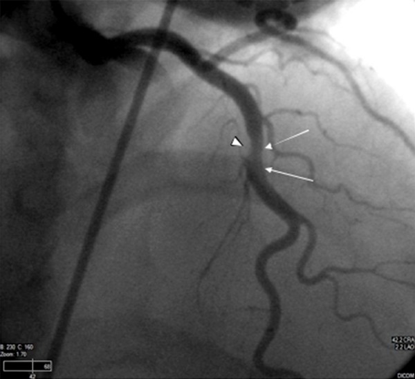
Figure 7.

LAD cranial view demonstrates eccentric narrowing (arrowhead) with differential luminal opacification at its midportion (white arrows). While there is no direct visualization of the intramural hematoma or intimal flap, these findings correlate well with the known dissection per prior CCTA.
