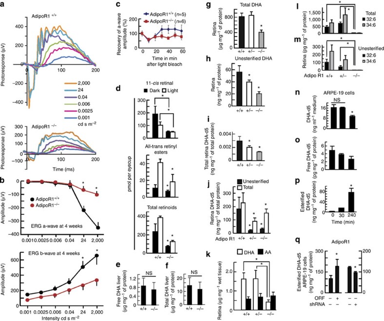
Figure 3. ERGs and visual cycle attenuated in AdipoR1−/− mice.
ERGs (3–4-week-old) before photoreceptor loss. (a) AdipoR1−/− had reduced ERGs. (b) AdipoR1−/− had attenuated a-/b-waves (a,b, n=4, AdipoR1+/+, n=6, AdipoR1−/−). (c) Impaired AdipoR1−/− recovery following 5-min bleaching light. Differences in AdipoR1+/+ and −/− a-waves demonstrated impaired recovery trend (n=5, AdipoR1+/+, n=6, AdipoR1−/− mice: 4–6-week old). (d) Impaired retinoid visual cycle (3–4-month old). (Dark-adapted mice 2,000 Lux, 10 min). 11-cis-retinal, all-trans-retinyl esters and total retinoids were diminished in AdipoR1−/− in light and darkness (n=4: both genotypes). Liver DHA is not altered in AdipoR1−/− mice. (e,f) Total and unesterified DHA showed no differences between 12-day AdipoR1+/+ and −/− (n=6 each). DHA uptake is reduced in AdipoR1−/− mice. (g,h) Total and unesterified DHA (20-day-old AdipoR1+/+, n=14, +/− n=31 and −/− n=7. Total DHA declined within AdipoR1−/−; unesterified DHA declined in AdipoR1+/− (30%) and AdipoR1−/− (60%). AdipoR1 deficiency reduced DHA uptake. (i) AdipoR1+/+ (n=6), +/− (n=20), and −/− (n=5) were injected ip with DHA-d5 (14 days), and retinas harvested (20 days). DHA uptake declined in AdipoR1−/−; intermediate DHA levels occurred in AdipoR1+/−, indicating a single allele cannot control DHA levels. (j) Eye cup cultures (n=5, all three genotypes, 20-days-old) incubated (4 h) with DHA-d5. AdipoR1 loss resulted in reduction of total DHA in AdipoR1+/− (60%) and −/− (30%) retinas. AdipoR1 selectively regulates DHA retinal uptake. (k) Total arachidonic acid (AA; 28-day-old mice) was similar in AdipoR1+/+ and −/−, while AdipoR1−/− DHA was decreased (75%). PC-associated VLC-PUFAs were reduced in AdipoR1−/−, with almost complete loss of total and unesterified 32:6 and 34:6 (l,m). (AdipoR1+/+, n=17; AdipoR1+/−, n=17; AdipoR1−/−, n=11). ARPE-19 cells incubated with DHA-d5 (100 nM) showed (n) media DHA-d5 decreased (50%), indicating DHA uptake, unesterified DHA declined (o) and DHA esterification increased (p), indicating DHA phospholipid incorporation. ARPE-19 cells incubated with DHA-d5 (240 min) and AdipoR1 overexpression enhanced DHA uptake and esterification; silencing decreased uptake and incorporation (q). (See AdipoR1 protein levels: Supplementary Fig. 1.) (n–q, statistical bars are s.e.m. of three for each condition. Experiments conducted three times. In all analyses, error bars represent s.e.m., and *P<0.05: t-test. NS=nonsignificant P value.)

