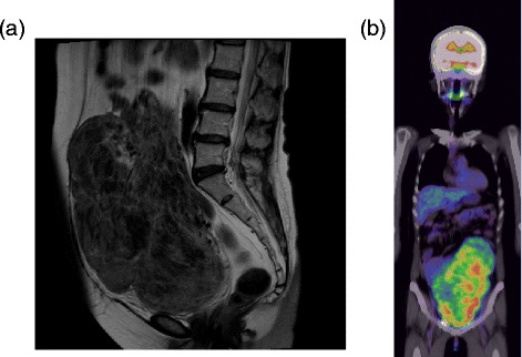
Figure 1.

Radiological images. (a) Pelvic magnetic resonance imaging, T2-enhanced sagittal section, showing an irregularly enlarged uterine tumor. (b) Positron emission tomography-computed tomography showing strong uptake associated with the uterine tumor.
