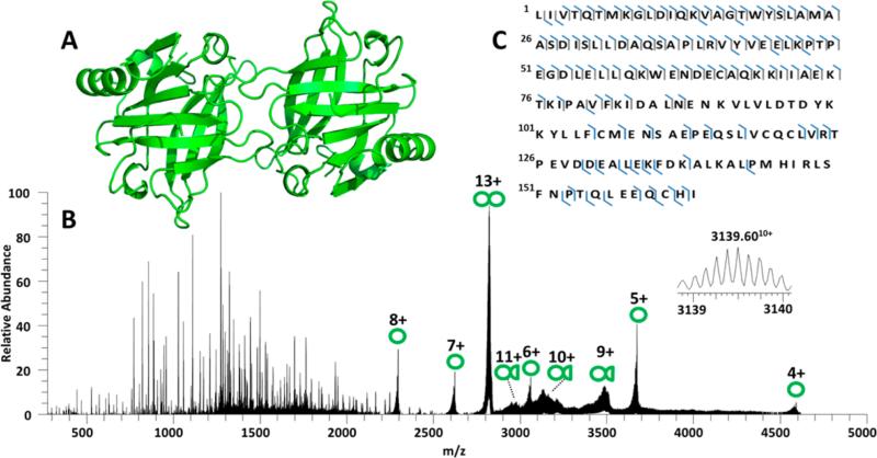
Figure 4.
(A) X-ray crystal structure of β-lactoglobulin (PDB: 1BSY). (B) UVPD mass spectrum of β-lactoglobulin dimer (13+). Single circles indicate intact monomers; double circles indicate dimers and circles with half circles are dimer fragments. The inset is an expansion of an identified a116 + β-lactoglobulin (10+) product ion. (C) UVPD sequence coverage map obtained for dimeric β-lactoglobulin (13+).

