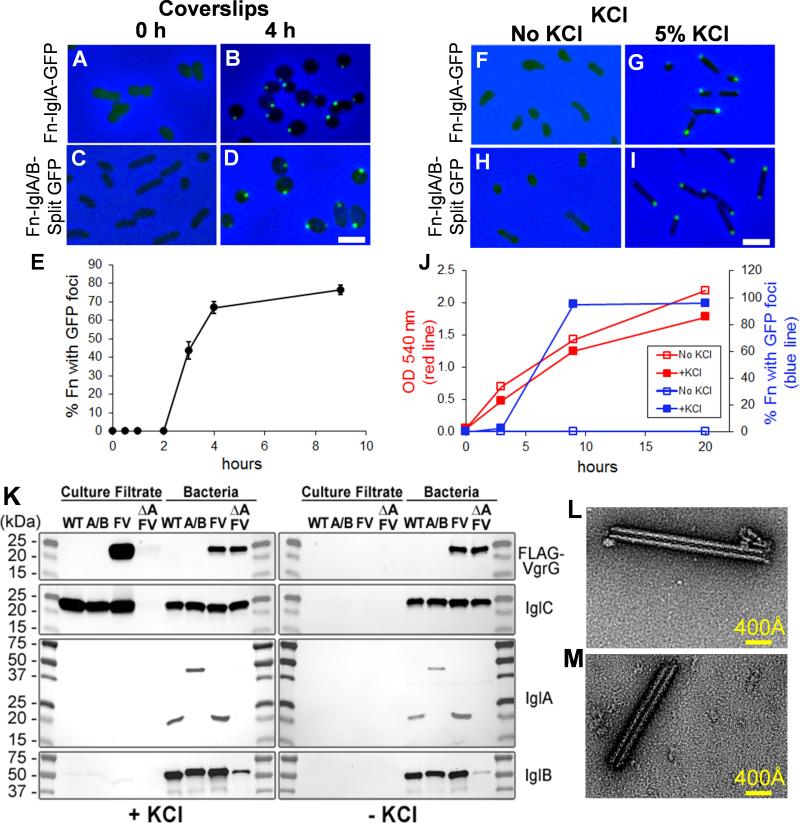
Figure 2. F. novicida assemble fluorescent foci in response to high KCl and placement beneath a coverslip and secrete T6SS effector proteins in response to environmental stimuli.
(A-E) Placement beneath a glass coverslip: Fn-IglA-GFP (A, B) and Fn-IglA/B-split GFP (C, D) in TSBC were placed beneath a silicone sealed coverglass and imaged immediately (Fn-IglA-GFP, A; Fn-IglA/B-split GFP, C) or after 4 h at room temperature (Fn-IglA-GFP, B; Fn-IglA/B-split GFP, D). Images shown are merged green fluorescence and phase contrast images, with phase contrast shown in blue. Bacteria exhibit either a diffuse weak fluorescence or no fluorescence immediately after placement beneath a glass coverslip (A, C), but exhibit intense fluorescence at their poles after 4 h at room temperature (Fn-IglA-GFP, B; Fn-IglA/B-split GFP, D). Scale bar, 2 μm. (E) Time course of formation of the fluorescent structures by Fn-IglA/B-split GFP after placement beneath a coverslip at room temperature.
(F-J) High KCl: Fn-IglA-GFP or Fn-IglA/B-split GFP were inoculated into TSBC at an O.D. 540 nm of 0.05 and grown at 37°C overnight to late exponential phase (O.D. 1.0 – 1.4) in the absence (Fn-IglA-GFP, F; Fn-IglA/B-split GFP, H) or presence of 5% KCl (Fn-IglA-GFP, G; Fn-IglA/B-split GFP, I). Bacteria exhibit only a weak diffuse fluorescence in the absence of KCl (F and H), but develop intense fluorescence at their poles in response to 5% KCl (G and I). Scale bar, 2 μm. See also Figure S1.
(J) Growth and kinetics of formation of fluorescent foci in Fn-IglA/B-split GFP in TSBC with and without 5% KCl. Fn-IglA/B-split GFP was inoculated into TSBC broth with or without 5% KCl at an optical density of 0.05 and grown at 37°C rotating at 250 rpm. O.D. (red lines) and percentage of bacteria with fluorescent foci (blue lines) in the presence (solid squares) or absence (open squares) of KCl were monitored over time.
(K) Secretion in response to high KCl: VgrG and IglC are secreted by F. novicida with intact T6SS growing in TSBC with (left), but not without 5% KCl (right). WT, wild-type; A/B, Fn-IglA/B-split GFP; FV, Fn-FLAG-VgrG; ΔA FV, Fn-FLAG-VgrG ΔiglA.
(L - M). Sheath like macromolecular structures purified from wild-type and IglA-GFP expressing F. novicida. F. novicida expressing native (L) or GFP-tagged (M) IgIA assemble similar sheath-like macromolecular structures. Negatively stained TEM images of density gradient fractions from F. novicida expressing wild-type IglA (L) or IglA-GFP (M) show rod shaped structures of variable length. See also Figure S2.

