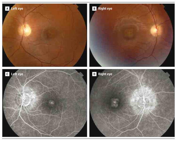
Figure 2. Clinical Presentation of the Disease in the Patient From Family MD-0312.
A and B, Funduscopy revealed normal retinal papilla and parenchyma, with normal vessels. A, Left eye; note changes at the foveal and perifoveal retinal pigment epithelium, including yellowish deposits, together with both nasal and inferior peripapillary atrophy. B, Right eye; note similar changes with the exception of the peripapillary atrophy, which was observed only at the nasal region. C and D, Fluorescein angiography demonstrated foveal hyperfluorescent dots and peripapillary hyperflourescence in both eyes. D, A higher extension of the affected area was observed in the right eye.

