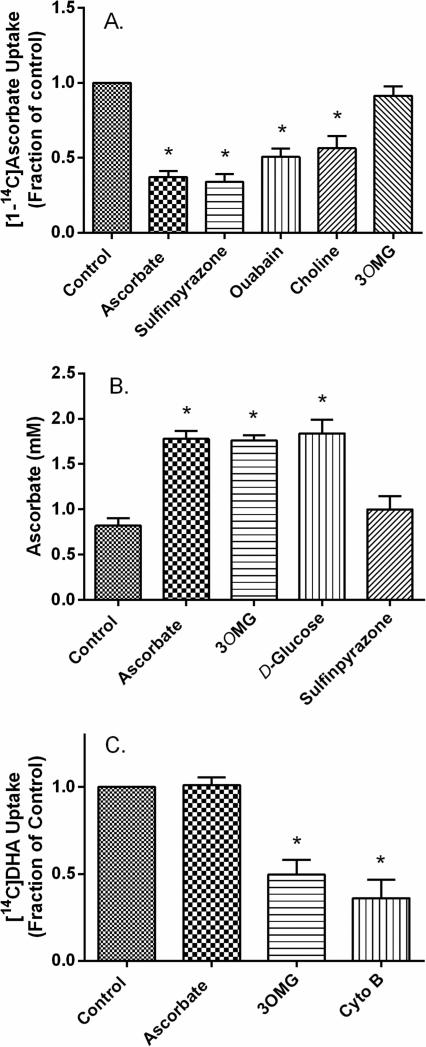
Figure 2. Inhibitor effects on radiolabeled ascorbate and DHA uptake.
Panel A. [1-14C]Ascorbate uptake. Using cultured pericytes, cold-stored medium was exchanged for KRH containing either 124 mM sodium chloride (first 5 bars) or 128 mM choline chloride instead of sodium (last bar). Cells in sodium-containing KRH were then incubated for 30 min with no additions (Control), with 1 mM ascorbate, with 2 mM sulfinpyrazone, with 300 μM ouabain, or with 30 mM 3-O-methylglucose (3OMG). After 30 min, 10 μM [1-14C]ascorbate was added to cells for an additional 30 minutes, and then cells were prepared for radioactive counting as described under Methods. Panel B. Uptake of unlabeled ascorbate. Cells cultured in fresh medium were untreated (first bar) or treated with 100 μM ascorbate, without or in addition to 3-O-methylglucose (3OMG, 30 mM), D-glucose (30 mM), or sulfinpyrazone (2 mM). After 30 min, cells were rinsed and harvested for assay of intracellular ascorbate. Panel C. [1-14C]DHA uptake. Cells cultured in cold-stored medium were treated for 30 min with the inhibitors and under the conditions noted in Panel A. Cytochalasin B (Cyto B, 10 μM) or 3-O-methylglucose (3OMG, 30 mM) were added where noted. [1-14C]DHA was prepared as described under Methods and its uptake at a final concentration of 10 μM was measured as described for [1-14C]ascorbate. Results in all panels are shown from 4-6 experiments, with those in A and C normalized in each experiment to an untreated control. “*” indicates p < 0.05 compared to the respective control.

