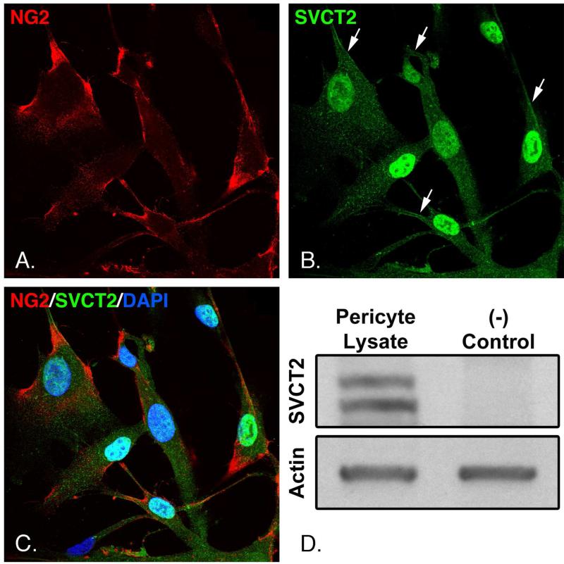
Figure 3. Presence of the SVCT2 in pericytes.
Panels A-C. Pericytes cultured on coverslips were fixed and probed with antibodies against the SVCT2 and NG2 as described under Methods. Cells were counterstained with DAPI to visualize nuclei and resolved by confocal microscopy at 600x magnification. Panel D. Pericyte lysates were subjected to gel electrophoresis and transferred to PVDF membranes as described under Methods. Membranes were probed with antibody against the SVCT2 (left lane), or with the same antibody pre-incubated with 5x its immunizing peptide as a negative control (right lane). The same membranes were probed for actin to confirm equal protein loading. Results shown are representative of 3 experiments.

