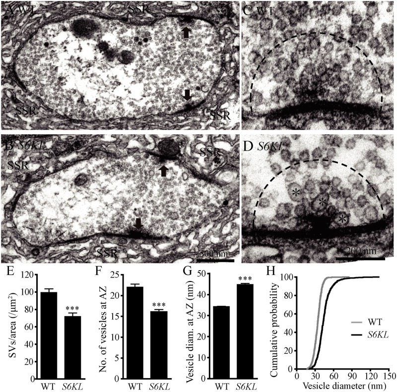
Fig 5. Fewer but larger synaptic vesicles at the active zone of S6KL mutant boutons.
(A–D) Ultrastructure of wild type (A and C) and S6KL 140 mutant (B and D) NMJ boutons. (A and B) Transmission electron micrographs of synaptic boutons. Arrows indicate active zones with T bars; SSR, sub-synaptic reticulum. Scale bar, 500 nm. (C and D) Higher-magnification images of active zones. Fewer and larger synaptic vesicles are present in S6KL 140 mutant active zones compared with wild type. Large synaptic vesicles are indicated by asterisks. Scale bar, 200 nm. (E–G) Quantification of the ultrastructural features: synaptic vesicle density in the whole presynaptic bouton area (E), the number (F) and diameter (G) of synaptic vesicles in a 200 nm radius around the T-bar. For the analysis of SV density, n = 73 and 46 boutons for wild type and S6KL 140, respectively. For the analysis of SV in a 200 nm radius around T-bar, n = 40 and 34 boutons for wild type and S6KL 140, respectively. ***p<0.001 by Student’s t-test; error bars indicate SEM. (H) Cumulative histograms of synaptic vesicle diameter indicating larger vesicles in S6KL 140 mutants.

