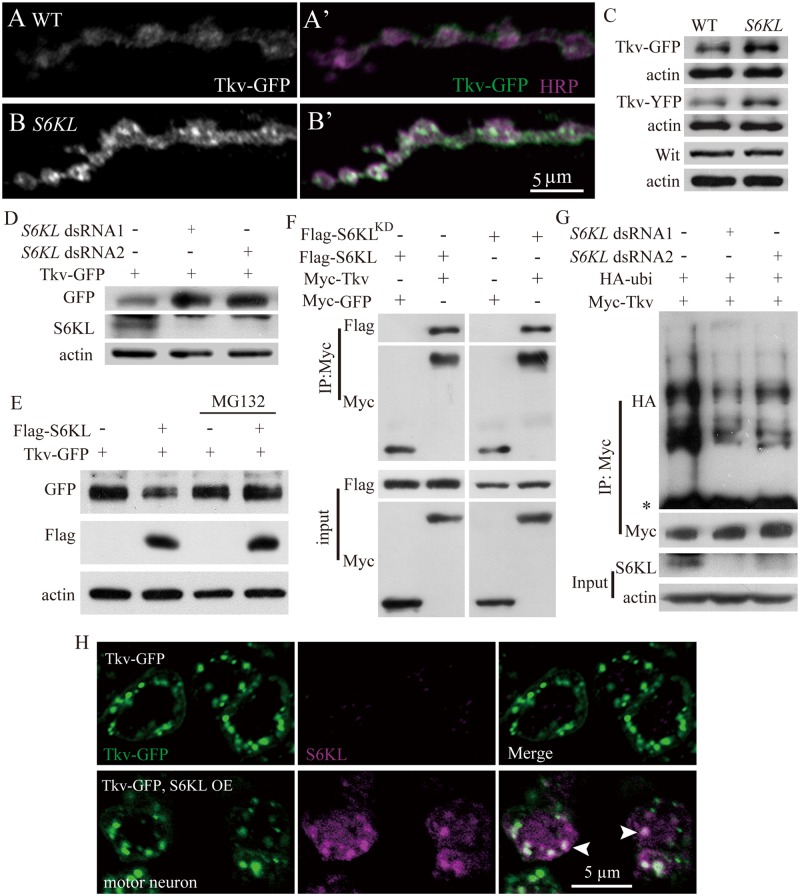
Fig 9. Negative regulation of Tkv protein level by S6KL via proteasomal degradation pathway.
(A–B′) Confocal images of NMJ 4 branches double-labeled with anti-GFP (green) and anti-HRP (magenta) in control (elav-Gal4/+; UAS-Tkv-GFP/+) and mutants (S6KL 140 elav-Gal4/S6KL 140 ; UAS-Tkv-GFP/+) showing elevated and punctate Tkv staining signals in S6KL 140 mutants. Scale bar, 5 μm. (C) Western results of total larval brain extracts probed with anti-GFP and anti-Wit antibodies. Actin was used as a loading control. (D) Tkv protein level was increased in S2 cells expressing a reduced level of S6KL. S2 cells were transfected with expression vector for Tkv-GFP and dsRNAs targeting two different sequences of the S6KL transcript. (E) Overexpression of S6KL suppresses Tkv protein level. S2 cells co-transfected with plasmids encoding Flag-S6KL and Tkv-GFP were untreated or treated with the proteasome inhibitor MG132 and subjected to western analysis with different antibodies. (F) Wild-type and kinase dead K193Q mutant (S6KLKD) S6KL interact with Tkv in S2 cells. S2 cells were co-transfected with expression vectors for Myc-Tkv or Myc-GFP and Flag-S6KL or Flag-S6KLKD. Cell lysates were subjected to immunoprecipitation with anti-IgG or anti-Flag and subsequently analyzed by western analysis. (G) Poly-ubiquitinated Tkv is decreased in S6KL—knockdown S2 cells. S2 cells were co-transfected with expression vectors for Myc-Tkv, HA-ubiquitin, and dsRNAs targeting two different sequences of the S6KL transcript. Asterisk denotes IgG. (H) S6KL and Tkv co-localize in the soma of motor neurons in the ventral nerve cord. Tkv-GFP alone (upper panels) or Tkv-GFP and S6KL (lower panels) were expressed under the control of the motoneuron specific OK6-Gal4. Scale bar, 5 μm.

