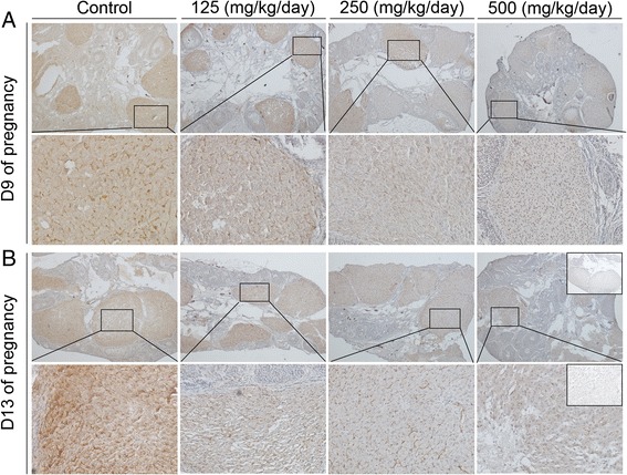
Figure 5.

Expression levels of CD31 protein detected by immunohistochemistry. (A, B) Representative images of ovaries on day 9 (A) and 13 (B) of pregnancy. Squared areas at the top (×40) are presented at higher magnification (×200) at the bottom. Inset is negative control.
