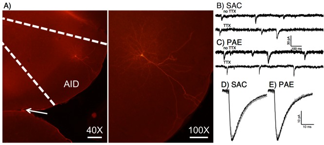
Fig 2. Sample layer II/III pyramidal neuron and spontaneous current recordings.

A) Example of biocytin-filled pyramidal neuron from cortical layer II/III. The internal solution for whole-cell patch-clamp electrophysiology contained biocytin to visualize patched neurons in paraformaldehyde fixed slices using fluorescence microscopy with streptavidin conjugated Cy3. A low magnification (40X) image is shown delineating agranular insular cortex, along with a higher magnification image (100X) to show neuronal morphology. The arrow in the 40X image points to the rhinal fissure. Scale bar 40X = 200 μm, scale bar 100X = 100 μm. B-E) Sample traces and average waveforms of sEPSCs and mEPSCs from AID layer II/III pyramidal neurons. Sample traces from saccharin exposed (B) and PAE (C) animals with sEPSC traces (no TTX) and mEPSC (TTX) traces are shown. Average waveforms for both sEPSCs and mEPSCs from saccharin exposed (D) or PAE (E) animals are also displayed. There were no significant effects of prenatal treatment condition on EPSCs.
