Fig. 1.
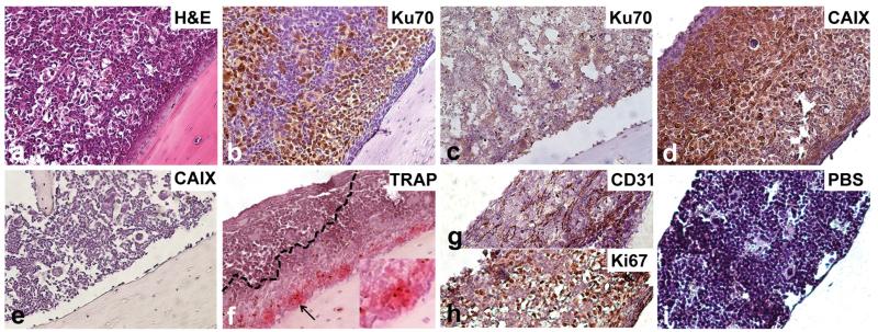
Immunophenotype of TSG bone metastases. Bone metastasis in femur of a mouse bearing case 1 TSG at passage 6. Photomicrographs (magnification ×20) showing H&E staining of bone marrow (a), immunohistochemistry for Ku70 (b) and CAIX (d), specific activity of TRAP, indicative of osteoclastic resorption capacity (f), and immunohistochemistry for CD31 (g), Ki67 (h), and negative control (i). Control bone stained with Ku70 (c) and CAIX (e) was negative. Insert in (f) shows a ×40 magnification of the area indicated by the black arrow

