Fig. 5.
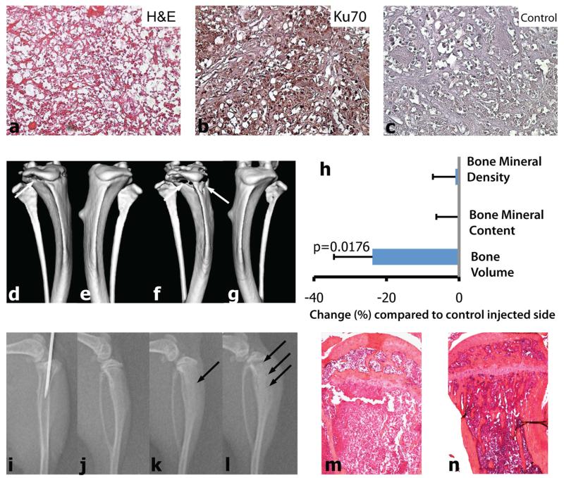
Intratibial injection of primary RCC cells results in bone osteolysis in mice. Precision-cut 300 lm thick tissue slices from case 1 TSG at passage six were digested into single cells with collagenase. Histology of frozen sections of precision-cut TSGs confirmed cancer. a H&E-staining. b Immunohistochemical staining for Ku70. c Negative control. The magnification for a-c is ×20. The correct position of the needle was confirmed with X-ray and single cells were injected into tibiae of mice (i). μCT analysis confirmed the advancing lytic appearance of the lesions: tumor-bearing right tibia 4- (d) and 6-(f) weeks after injection of primary cancer cells; e and g, corresponding left tibia with no tumor. h Volumetric bone analysis with μCT showed reduction in bone volume of the tumor-bearing tibiae at 6 weeks after cancer cell inoculation compared to control injected side (n = 3). Consecutive X-ray images (j, day 0, i.e., before injection, and k and l, 3- and 5-weeks after injection, respectively) revealed development of advancing lytic lesions (arrows in k and l). m H&E-staining of cross-section of tumor cell-injected tibia revealed destruction of trabecular bone and thinning of cortical bone compared to HBS-injected control tibia (n). Magnification for m and n is ×4

