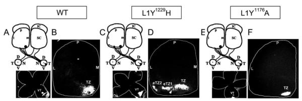
Figure 2. Normal retinocollicular mapping of L1Y1176A mutant mice.

A-B. Injection of DiI into the peripheral ventro-temporal (VT) retina of WT mice (P10) labeled a single termination zone (TZ) in the anteromedial superior colliculus (SC) at P12.
C-D. DiI injection into the ventro-temporal retina of homozygous L1Y1229H mutant mice showed multiple laterally-shifted ectopic TZs (eTZs) in the anterior SC.
E-F. DiI injection into the ventro-temporal retina of homozygous L1Y1176A mutant mice showed only a single normal TZ in the anteromedial SC.
The position of injections is shown in flat mounts of the retina below each scheme. (L, lateral; M, medial; A, anterior; P, posterior; D, dorsal; V, ventral; N, nasal; T, temporal; IC, inferior colliculus)
