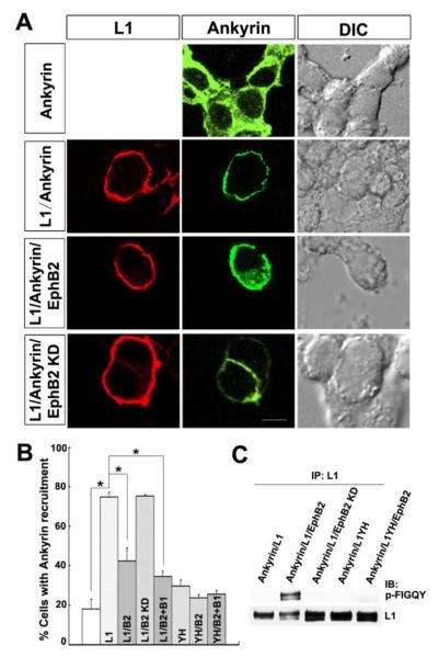
Figure 3. EphB2 kinase inhibits recruitment of ankyrin to cell membrane of L1-expression cells in a cytofluorescence assay.
A. Immunofluorescence staining for L1 in the plasma membrane of transfected HEK293 cells (left column, red) and EGFP-ankyrinG fluorescence (middle column, green) showed that EGFP-ankyrinG had a cytoplasmic distribution when expressed alone (Ankyrin), while L1 expression recruited EGFP-ankyrinG to the cell membrane (L1/Ankyrin). EGFP-ankyrin remainedG cytoplasmic when L1 was co-expressed with EphB2 (L1/Ankyrin/EphB2) but not with EphB2 KD (L1/Ankyrin/EphB2 KD).
Right column shows differential interference contrast (DIC) images. Scale bar=10 um
B. Quantification of percentage of cells showing ankyrin recruitment to the plasma membrane demonstrated that L1 increased ankyrin recruitment to the membrane in cells co-expressing L1 and ankyrinG compared to ankyrinG alone (L1/ankyrin: 75 ± 2%; ankyrin: 18 ± 5%; one-way ANOVA, Tukey’s post-hoc test, *p<0.001).
Recruitment decreased in L1/ankyrin/EphB2 expressing cells (42 ± 6 %) compared to L1/ankyrin (*p<0.001). There was no decrease of recruitment in L1/ankyrin/EphB2 KD (75 ± 0.5%) compared to L1/ankyrin expressing cells. EphrinB1 treatment (35 ± 3%) reduced ankyrin recruitment to a small extent in L1/ankyrin expressing cells (*p<0.001). No significant difference was detected between ankyrin alone and negative controls of ankyrin/L1Y1229H (29 ± 3%), ankyrin/L1Y1229H/EphB2 (24 ± 1.5%) or ankyrin/L1Y1229H/EphB2 + ephrinB1 (26 ± 2%) (p>0.05).
(Labeling on bars indicate cells transfected with ankyrinG alone (first bar), L1/ankyrin (L1), L1/ankyrin/EphB2 (L1/B2), L1/ankyrin/EphB2 KD (L1/B2 KD), L1/ankyrin/EphB2+ephrinB1 (L1/B2+B1), L1Y1229H/ankyrin (YH), L1Y1229H/ankyrin/EphB2 (YH/B2), L1Y1229H/ankyrin/EphB2+ephrinB1 (YH/B2+B1).
C. L1 is phosphorylated by EphB2 at the FIGQY motif in ankyrin-expressing HEK293 cells.
Under the conditions of the ankyrin recruitment assay, EGFP-ankyrinG expressing HEK293 cells were co-transfected with L1, L1/EphB2, L1/EphB2 KD, L1YH, or L1YH/EphB2. L1 was immunoprecipitated from equal amounts of cell lysates (500 μg) and immunoblotted with p-FIGQY antibodies, then blots were stripped and reprobed with L1 antibodies. EphB2, but not EphB2 KD, induced L1 phosphorylation at FIGQY, while L1YH was not phosphorylated by EphB2.

