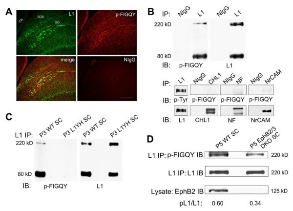
Figure 4. L1 FIGQY motif is phosphorylated in retina ganglion cell axons in the superior colliculus.
A. In WT mice (P7), immunofluorescence staining for p-FIGQY (red) in sagittal sections through the SC co-localized in part with L1 (green) in fibers within the superficial retino-recipient layers (SGS and SO) where RGC axons track, as indicated in merged images. P-FIGQY staining was also seen in some fibers in the retino-recipient layers, and in deeper layers of the SC. No staining was detected with normal rabbit IgG. Scale bar = 200 μm. (SGS, stratum griseum superificiale; SO, stratum opticum; Ctx, cerebral cortex)
B. p-FIGQY antibody recognized full length L1 (220 kDa) and its principal C-terminal cleavage fragment (80 kDa) in the SC, as shown by immunoblotting in L1 immunoprecipitates from WT SC lysates (P3) (upper panel). The p-FIGQY antibody also recognized NrCAM to a lesser extent but not CHL1 or Neurofascin (NF) immunoprecipitated from P3 SC (lower panel).
No bands were detected in normal mouse IgG precipitates in any sample.
C. p-FIGQY antibody did not recognize L1 immunoprecipitated from L1YH mutant SC (P3).
D. The relative level of p-FIGQY in full length 220 kDa L1 was decreased in L1 immunoprecipitated from EphB2/B3 double null mutant SC (P5) compared to WT SC. EphB2 protein was undetectable in EphB2/B3 double mutant SC.

