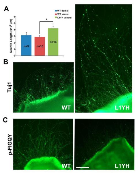
Figure 5. Increased outgrowth of retinal axons from L1Y1229H mice in explants cultures from ventral retina.
A. Quantification of mean neurite length of TuJ1-positive neurites extending from ventral retinal explants demonstrated that outgrowth of RGC axons from L1Y1229H mice was significantly greater than that of WT mice (t-test, *p<0.001). There was no significant difference in mean neurite length between WT dorsal and WT vental retinal explants.
n = number of mice of each genotype.
B. Ventral retina explants from L1Y1229H mutant mice showed RGC axons extending far from the explants edge in addition to some of comparable length compared to WT, indicated by immunostaining with neuronal marker TuJ1.
C. Immunostaining of ventral retinal explant cultures from WT and L1Y1229H mutant mice with p-FIGQY antibody showed no staining of L1YH mutant axons, while strong p-FIGQY staining was seen in WT axons.
Scale bar = 100 μm in B-C.

