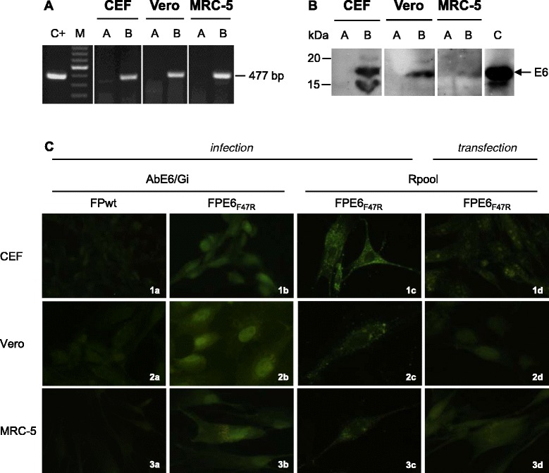
Figure 2.

Expression of the E6 F47R in replication-permissive avian and replication-restrictive mammalian cells. RT-PCR was used to amplify the 477-bp E6-specific transcripts in all of the cell types (A, lanes B). FPwt-infected cells were used as negative controls (A, lanes A) and the plasmid pFPE6F47R as a positive control (C+). M; 100-bp ladder. Western blotting was used to reveal the presence of a 19-kDa protein in cells infected by FPE6F47R (B, lanes B) using the rabbit AbE6/Mu polyclonal antibody. FPwt-infected cells (B, lanes A) and the E6 protein (B, lane C) were used as a negative or positive controls, respectively. By immunofluorescence (C), specific staining was detectable in all cell lines after infection with FPE6F47R (C; 1b, 2b, 3b and 1c, 2c, 3c) or transfection with pDNAE6F47R (C; 1d, 2d, 3d) recombinants, using either the mouse AbE6/Gi or the rabbit Rpool polyclonal antibodies. FPwt-infected cells were negative (C; 1a, 2a, 3a), as well as the pcDNA3-transfected cells, as expected (data not shown).
