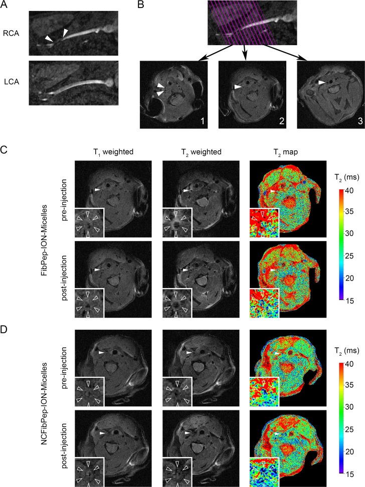
Fig 3. In vivo MRI.
(A) Images of right and left carotid arteries (RCA & LCA) reconstructed from the 3D time of flight (TOF) image. The thrombus can be observed in the RCA just proximal of the bifurcation (arrowheads). Presence of blood flow in the thrombosed carotid was confirmed for all animals. (B) On the TOF image 13 parallel slices were planned perpendicular to the RCA. T1-weighted images from three slices are shown: (1) distal from the bifurcation, (2) in the thrombus and (3) proximal to the thrombus (arrowheads: RCA). (C, D) Pre- and post-injection T1- and T2-weighted images and T2 maps of (C) FibPep-ION-Micelles and (D) NCFibPep-ION-Micelles (arrowheads: RCA). The insets show magnifications of the RCA (open arrowheads: outer vessel wall).

