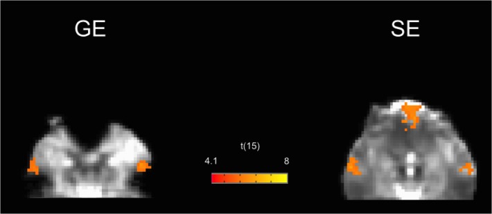
Fig 4. GE and SE group connectivity maps for the default mode network in the inferior brain regions.
The map is superimposed on the Talairach transformed EPI images of one subject. The signal void is clearly visible in the GE images, with the corresponding loss of functional contrast in the ventromedial prefrontal node. Both raw signal and functional contrast are instead recovered in SE images.

