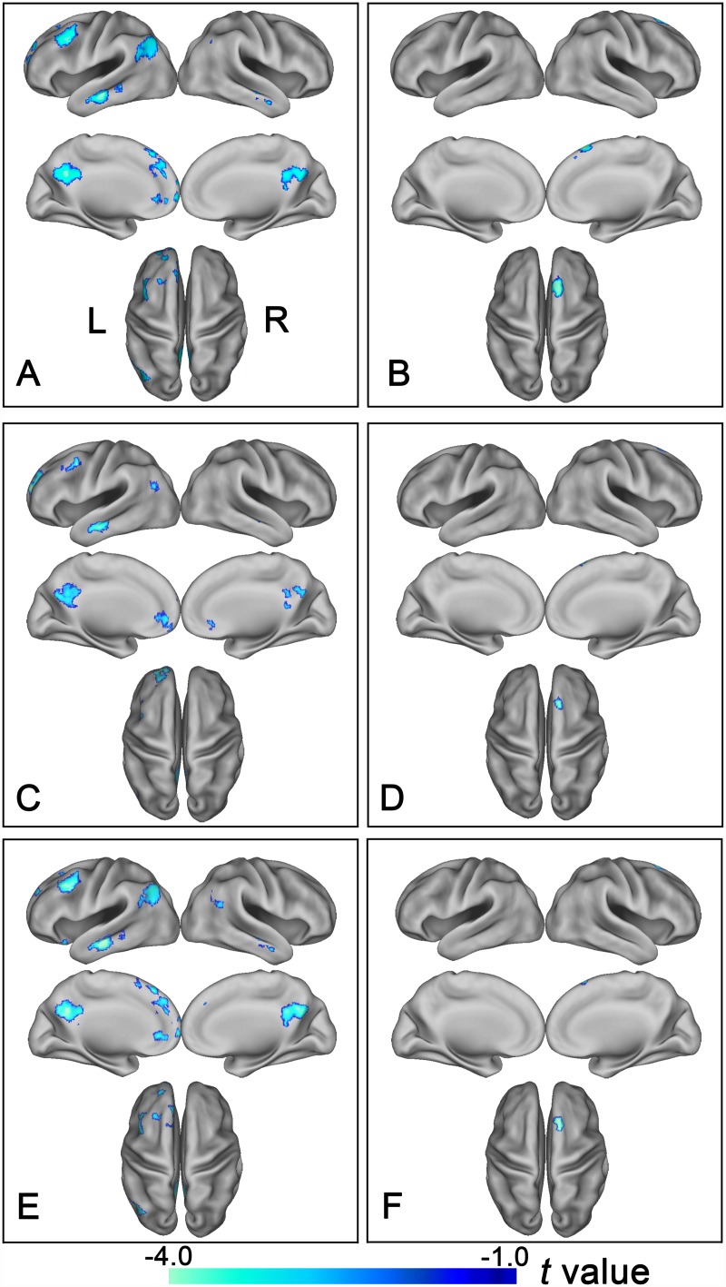
Fig 3. Altered rsFC of the bilateral lateral subregions of the frontal pole in schizophrenia patients.
Brain regions exhibit significantly reduced rsFC with the left FPl (A, C) and the right FPl (B, D) in schizophrenia patients. (A, B) show results derived from the method of the maximal probability map and (C, D) demonstrate results derived from the method that using spherical regions of interest as seed regions. Of note, the between-group differences in the rsFC of the bilateral lateral subregions of the FP exhibited highly similar patterns between with (E and F) and without (A, B) correcting GM volume. The statistical threshold was set at p<0.05, FDR correction, two-tailed and cluster size>30 voxels. FPl, lateral subregion of the frontal pole; FP, frontal pole; L, left, R, right.

