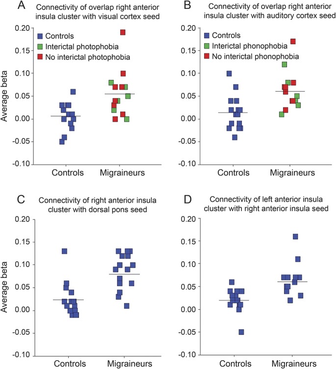
Figure 3. Subject-specific scatterplots of connectivity metrics within anterior insular clusters.
Each square represents the mean β value within the insular cluster of interest for one subject; horizontal line indicates group mean. Subject-specific scatterplots are shown for connectivity within the overlap right dorsal anterior insula cluster (figure 1C, z = 22) from the (A) calcarine cortex–seeded and (B) auditory cortex–seeded maps, (C) within the right anterior insula cluster from the dorsal pons–seeded map (figure 2A), and (D) within the left anterior insula cluster from the right anterior insula–seeded map (figure 2B, z = −8). In A and B, migraineurs are further color-coded according to whether they reported interictal photophobia or phonophobia, respectively.

