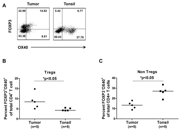
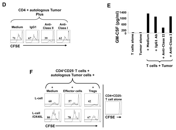
Figure 5. Triggering OX40 enhanced effector T cell function and blocked Treg suppression in FL.
A–C) Expression of OX40 was determined by flow cytometry on Foxp3+ (A,B) and Foxp3- (A,C) CD4+ T cells in FL tumors and tonsils. Representative data (A) and data from 5 FL tumors and tonsils (B,C) are shown. Horizontal bars indicate median values for each group. P values were calculated by Wilcoxon rank-sum test. D and E) CFSE-labeled (D) or unlabeled (E) intratumoral CD4+CD25- effector T cells were cultured with autologous CD3-depleted FL tumor-derived antigen-presenting cells in the presence or absence of anti-MHC Class I or II antibodies or isotype control antibody. Proliferation was assessed by CFSE dilution after 4 days (D) and GM-CSF production was measured in the culture supernatants after 24 hours (E). F) Effector T cells were stimulated as in panel D and cultured with parental L cells or L cells expressing OX40 ligand in the presence or absence of autologous intratumoral Tregs. Numbers shown in each histogram indicate the percentage of proliferating effector T cells (D, F).


