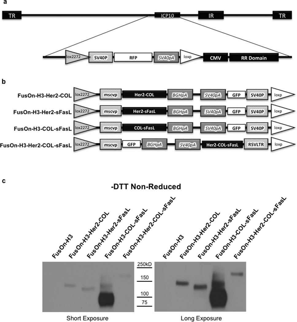
Figure 3. Construction of FusOn-H3 recombinant viruses and secretion of chimeric sFasL molecules following infection.

(a) Schematic representation of FusOn-H3-RFP virus genome. The modified region adjacent to the ICP10 gene locus is enlarged showing the lox2272/loxp flanked RFP cassette. TR, terminal repeat; IR, internal repeat; ICP10, infected cell protein 10 gene; loxp and lox2272, cre recombinase binding sites; SV40P, SV40 promoter; RFP, red fluorescent protein gene; SV40pA, SV40 polyadenylation sequence; CMV, cytomegalovirus promoter; RR1, ribonucleotide reductase carboxyl terminus. (b) PCR2.1 donor plasmid inserts for recombinant FusOn-H3 construction. Mscvp, murine stem cell virus promoter; BGHpA, bovine growth hormone polyadenylation signal; GFP, green fluorescent protein; RSVLTR, rous sarcoma virus long terminal repeat. (c) Western blot confirmation of sFasL chimeric molecule secretion by recombinant FusOn-H3 virus infected cells. HCT116 cells were infected at an MOI of 1. Media was collected 24 hours post infection and equal volumes subjected to Western blot under non-reducing conditions. Proteins were detected using an HA tag antibody and are shown at two different exposure times for clarity. Data are representative of two independent experiments.
