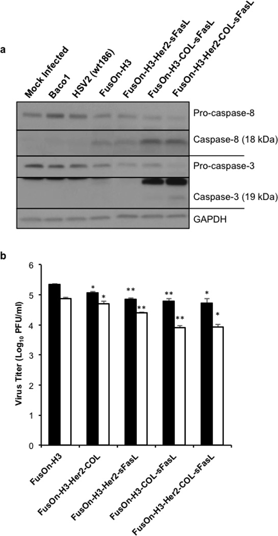
Figure 4. Arming of FusOn-H3 with apoptosis activators increases caspase activation without severely effecting replication.

(a) Western blot of caspase-8 and caspase-3 cleavage following recombinant virus infection. HCT116 cells were infected at an MOI of 0.1, cells collected 30 hours post infection, and lysates subjected to Western blot analysis. Caspase-8 and capsase-3 antibodies were used to detect specified cleavage products. GAPDH was used as a loading control. Data shown is representative of two independent experiments (b) Viral titer of FusOn-H3 and recombinant viruses expressing chimeric sFasL molecules. HCT116 cells were infected in triplicate with designated virus at an MOI of 1. Cells were collected 24 (black bars) and 48 hours post infection (white bars) and total infectious virus quantified through titration in Vero cells. *p< 0.01; **p<0.001 compared to respective 24 or 48 hour FusOn-H3 titer according to student’s T test. Error bars represent mean ±SEM.
