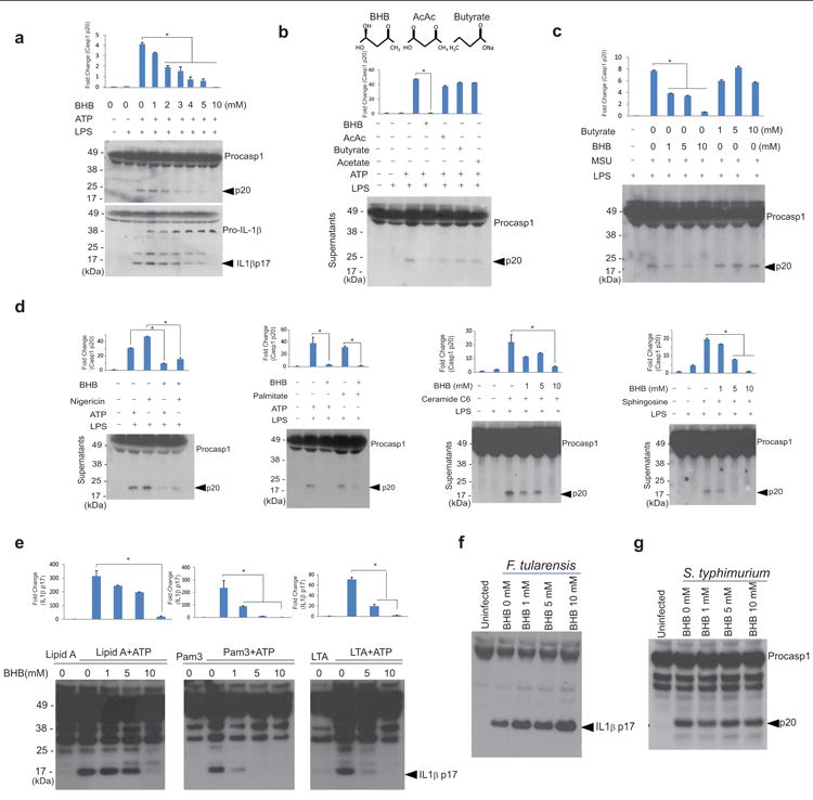
Figure 1. BHB specifically inhibits the NLRP3 inflammasome.

(a) Representative Western blot analysis of caspase-1 (active subunit p20) and IL-1β (active p17) in the supernatant of BMDMs primed with LPS for 4 hours and stimulated with ATP for 1 hour in the presence of various concentrations of D-BHB. (b) Western blot analysis of caspase-1 activation in BMDMs stimulated with LPS and ATP and treated with BHB (10mM), butyrate (10mM), acetoacetate (10mM) and acetate (10mM). (c) Western blot analysis of caspase-1 activation in LPS- primed BMDMs stimulated with MSU and treated with butyrate and D-BHB (d) nigericin (10 μM) for 1h, palmitate (200μM) for 24h, C6 ceramide for 6h (80μg/ml), and sphingosine (50 μM) for 1hour. (e) Western blot analysis of IL-1β activation (active subunit p17) in BMDMs primed with TLR ligands lipid A, Pam3-CSK and LTA for 4h and stimulated with ATP and increasing doses of D-BHB for 1h. Active IL1β (p17) was analysed in supernatants by western blot. BMDMs were infected with (f) F. tularensis and (g) S. typhimurium and treated with different doses of BHB and IL1β activation (p17 active form) was analyzed. Data are expressed as mean ± S.E.M (*P < 0.05) from cells derived from twelve (a-d) or six (e), three (f, g), mice with each independent experiment each carried out in triplicate (a-d, e) and duplicate (f g). All bar graphs in (a-e) represent quantitation of p20 caspase-1 band intensity as fold-change by normalizing to inactive p48 procaspase-1, or p17 IL-1β band intensity as fold change by normalizing to inactive p37 pro-IL-1β. The differences between means and the effects of treatments were determined by one-way ANOVA using Tukey's test.
