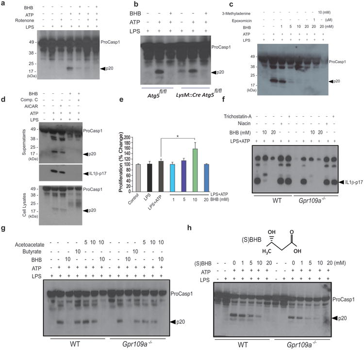
Figure 2. BHB inhibits the NLRP3 inflammasome independently of Gpr109a and starvation-regulated mechanisms.

(a) Western blot analysis of caspase-1 activation in LPS- primed BMDMs treated with rotenone (10μM), ATP (5μM) together with BHB (10mM). (b) Western blot analysis of caspase-1 activation in BMDMs derived from control Atg5fl/fl and LysM:Cre Atg5fl/fl mice primed with LPS and stimulated in presence of ATP and BHB (10mM) (c) The BMDMs were primed with LPS and pretreated with 3MA and epoxomicin for 30min and stimulated in presence of ATP and BHB. The caspase-1 activation was measured by immunoblot analysis. (d) Western blots of caspase-1 and IL-1β activation in LPS-primed BMDM stimulated with ATP and BHB (10mM) in presence of AMPK activator (AICAR, 2mM) and AMPK antagonist Compound C (25 μM). (e) Proliferation of BMDMs in response to increasing concentrations of BHB. (f ,g) Western blot analysis of caspase-1 and IL-1β activation in BMDMs from control and Gpr109a deficient mice activated with LPS and ATP and co-incubated with TSA (50nM), niacin (1mM), butyrate (10mM), acetoacetate (10mM) and BHB (10, 20mM) (h) Western blot analysis of caspase-1 activation in BMDMs of WT and Gpr109a-/- mice treated with LPS for 4h and stimulated with ATP in presence of BHB chiral enantiomer (S) BHB for 1h. Data are expressed as mean ± S.E.M (*P < 0.05) from cells derived from six (a) 4 (b) ten (c, d, e) and four (f-h) mice with each independent experiment each carried out in triplicate. Due to space limitations the quantitation of p20 caspase1 and p17 IL1β band intensity from each experiment are presented in the Supplementary Fig. 2A. The differences between means and the effects of treatments were determined by one-way ANOVA using Tukey's test.
