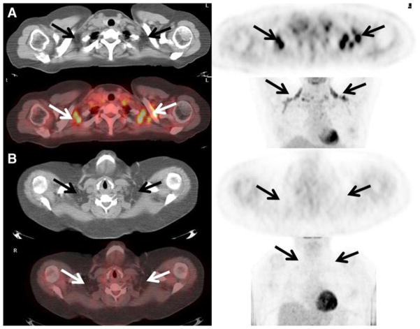
Fig. 1.
a Active brown adipose tissue detected on FDG PET/CT. A 48-year-old female with a history of endometrial carcinoma underwent FDG PET/CT to evaluate for recurrent disease. Foci of intense FDG uptake are seen fusing to fat in the supraclavicular regions consistent with the presence of active BAT (arrows). Her blood glucose level just prior to the administration of FDG was 72 mg/dL. Her body mass index was 26. b No active brown adipose tissue detected on FDG PET/ CT. A 50-year-old female with a history of non-Hodgkin’s lymphoma underwent FDG PET/CT for re-staging purposes. No increased FDG uptake was seen localizing to fat on the CT scan suggesting the absence of metabolically active BAT (arrows). Her blood glucose level just prior to the administration of FDG was 127 mg/dL. Her body mass index was 49.

