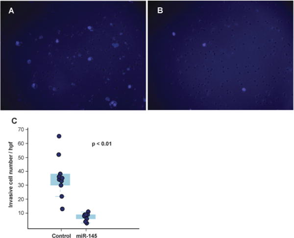
Fig. 4.

Decreased invasiveness of pancreatic cancer cells mediated by miR-145 in matrigel invasion assays. Cell nuclei of MiaPaCa-2 were stained with DAPI and are visualized as light purple dots by fluorescence microscopy. (A) Forty-eight hours after scrambled RNA transfection. (B) Forty-eight hours after miR-145 transfection. (C) Quantitation of invasive cancer cells depicted as a box-dot plot.
