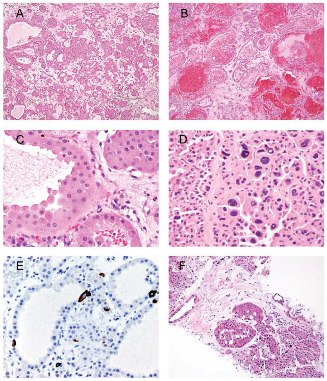
Figure 1.
A, Classic appearance of oncocytoma with small solid nests with myxoid or hyalinized background. B, Tubular architecture of oncocytoma with hemorrhage. C, Classic cytology of oncocytoma with abundant eosinophilic cytoplasm; indistinct cell borders; small, uniform nuclei; and prominent nucleoli. D, A cluster of degenerative-type atypia seen in oncocytoma. E, Scattered cytokeratin 7 immunoreactivity in oncocytoma. F, Biopsy of an oncocytic renal neoplasm consistent with oncocytoma (hematoxylin-eosin, original magnifications ×100 [A, B, and F], × 600 [C], and ×400 [D]; original magnification ×400 [E]).

