
Figure 2.
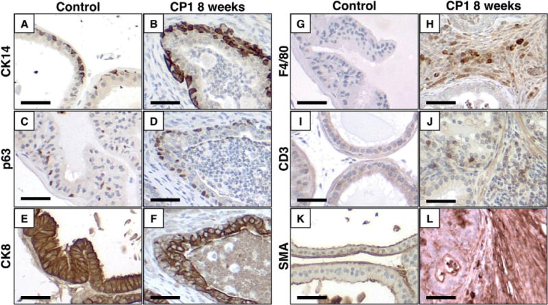
CP1-infected prostates are hyperplastic and infiltrated by immune cells. Representative images of immunohistochemistry comparing saline-treated controls and CP1-infected prostates 8 weeks after infection demonstrate hyperplasia of basal cells (CK14, A, B; p63 C, D), luminal cells (CK8, E, F), and thickened stroma with disruption of the smooth muscle actin-positive layers (SMA, K, L). Inflamed prostates show increased infiltration of macrophages (F4/80, G, H) and T cells (CD3, I, J). Scale bar =50 μm.