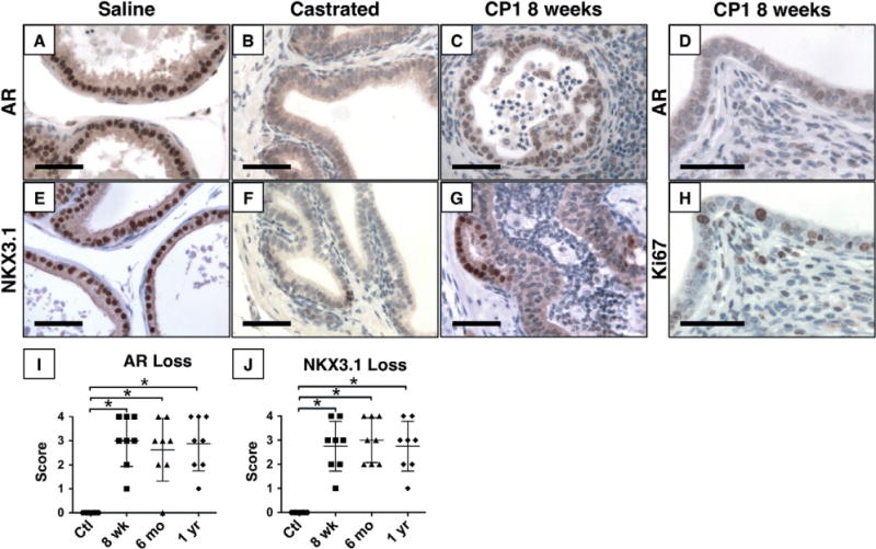
Figure 5.

Diminished AR expression and signalling in inflamed epithelium. Immunohistochemistry for AR (A–D) and NKX3.1 (E–G), an androgen regulated protein, shows strong nuclear expression in normal glands (A, E), but significantly decreased expression of both proteins 2 weeks after castration (B, F). Expression of both proteins is decreased to castrate levels in inflamed epithelium 8 weeks post-inoculation (C, G). Despite decreased AR expression, inflamed epithelium remains highly proliferative with numerous Ki67-positive nuclei (D, H). Quantification of the percentage of positive nuclei shows significant loss of AR (I) and NKX3.1 (J) expression (p <0.001, n =8 per group). Scale bar =50 μm.
