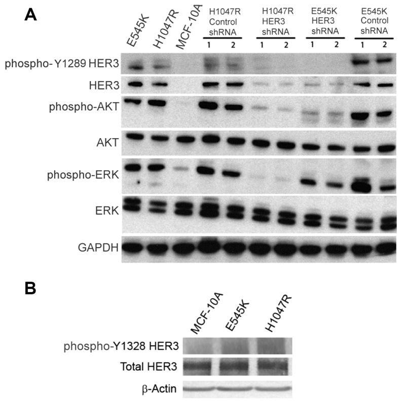
Figure 3.
HER3 knock down results in decreased Akt phosphorylation in MCF-10A PIK3CA mutant E545K and H1047R knock in cell lines, and decreased Erk phosphorylation in H1047R cells. (A) Western blot demonstrating levels of phosphorylated HER3 (Y1289), total HER3, phosphorylated Akt (Ser-473), total Akt, phosphorylated Erk (Thr-202/Tyr-204), total Erk, in MCF-10A cell lines, derivatives and controls. GAPDH is shown as a loading control. (B) Western blot demonstrating levels of phosphorylated HER3 (Y1328) and total HER3 in MCF-10A cells and PIK3CA E545K and H1047R knock in cell lines. GAPDH is shown as a loading control.

